Free Royal Mail 24 Tracked Delivery - Spend £10+
 Support 01904 789559 - 20+ Years Expertise
 Rated 4.9 out of 5 on Trustpilot


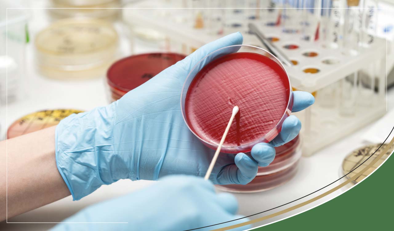
Urine Analysis, Microscopic & Chemical Examination

What is Urine?
Blood passes through the kidneys, and next urine is formed and excreted. Urine is made up of substances that are not used or needed by our cells, so they are the leftovers of metabolic processes (e.g., urea). The blood is first filtered and all small molecules, including both nutrients and wastes, enter a nephron. There are about 1,000,000 nephrons in each human kidney and nephrons are the active part of the kidney that produces urine while removing waste and excess substances from the blood. The nutrient molecules and some salts and water are reabsorbed back into the blood, while unwanted substances remain within the nephron to become a part of the urine.
Urinalysis
A urinalysis is, as the name suggests, is an analysis of the urine. A sample of 30-60mls of your urine is needed for urinalysis.
Your GP may order urinalysis in a lab to test for kidney, metabolic disorders and urinary tract infections. If you are menstruating, close to your period or are taking any diuretics, it is best to tell your GP as they may wish to postpone obtaining a sample. It is also best to avoid strenuous exercise or eating any foods that could colour your urine before the test - e.g. beetroots, blackberries or rhubarb.
Urinalysis may be needed for a number of reasons, for example:
- If you are experiencing pain in your lower abdomen or back, blood in the urine (either visible or found on a urine dipstick test), if you are experiencing pain when urinating or an increase in frequency of urination
- Routine medical examinations such as yearly check-ups, admission to hospital, screening for diabetes, kidney disease, hypertension, liver disease
- To evaluate new pregnancy
- To monitor progression and response to treatment of diseases such as diabetes, kidney impairment/disease/stones, lupus
- To determine the cause of bladder infections.
It is recommended to obtain a mid-stream sample in a clean, sterile tub (typically provided by your healthcare professional). You will be given instructions as to what time of day to collect the sample (for example, first urination of the morning). If the sample needs to be kept for more than an hour before it is collected and sent to the lab, it must be stored in a fridge (but for no longer than 24 hours).
There are three testing phases in a complete urinalysis: the Visual Examination, the Chemical Examination and the Microscopic Examination.
The Visual Examination
In the laboratory, the technician will check for clarity, colour and concentration of the urine.
Colour:
Sometimes the urine will be affected by diet - e.g. foods like beetroot can colour the urine red. Other times, red-coloured urine may indicate the presence of blood, suggesting disease or damage to the urinary system. It is important to also rule out blood-contaminated urine from menstruation or haemorrhoids. Normal urine colour usually ranges from a pale yellow colour to a little darker yellow first thing in the morning when urine is more concentrated. A darker amber colour suggests dehydration and that you need to drink more water, and very dark orange could be an indicator of jaundice/bile duct disorder or liver disorder.
Clarity:
The technician will review the urine also on its clarity - whether it is cloudy, turbid, clear. Healthy urine can still be cloudy or turbid if contaminated with substances such as male reproductive fluid or prostatic fluid, skin cells, asymptomatic urine crystals, mucus or personal hygiene products. However, cloudiness could also be an indicator of a health condition or infection since it can also be caused by the presence of bacteria or red or white blood cells in the urine.
The visual examination is only part of the story in urinalysis, to identify which substances are present, it is necessary to perform further chemical and microscopic tests on the urine.
The Chemical Examination
The chemical examination is usually performed in the laboratory using urine test strips with rows of test pads called reagents. Each pad on the urine test strip contains a different chemical that will change colour to indicate the presence of specific substances in your urine. The colour indicates whether a small or large amount of the substance has been detected. The results are often read by the technician; however, sometimes the reading is automated to prevent errors of interpretation and timing since each pad colour needs to be analysed at the correct time (ranging from a few seconds to minutes dependent on the reaction it is detecting) to ensure accurate results.
What does the chemical examination test for?
The chemical examination can test for a range of indicators:
- Protein
- Blood (Haemoglobin)
- pH Level
- Leukocytes
- Nitrates
- Bilirubin
- Urobilinogen
- Ketones
- Glucose
- Specific Gravity
Protein
Protein in the urine is usually measured by testing for the presence of albumin, but there is a range of alternative protein tests that can be used. High protein levels (proteinuria) can be an early indicator of kidney disease. Detection of protein in the test pad may also be an indicator of urinary tract infection/inflammation or injury such as damage to the prostate, bladder or urethra. Proteinuria could also indicate conditions such as bladder or kidney stones, multiple myeloma, or any condition that destroys red blood cells such as haemolytic anaemia. It is often a temporary phenomenon that disappears after dealing with an infection.
Blood (Haemoglobin)
Haemoglobin is the protein found in red blood cells and carries oxygen. The urine normally contains a small amount of haemoglobin which does not show up on the test, but a higher amount (haemoglobinuria) will show up as a positive test result. It is important that instructions are followed when collecting the sample, so that is not contaminated through menstrual blood or blood from haemorrhoids. A slight increase from the norm in haemoglobin can be significant in terms of potential causes such as a urinary tract infection, kidney disease, trauma, strenuous exercise. Smoking or certain medications can also show an increase.
pH Level
The urine must be tested within a few hours of collection to avoid skewing the results for the pH test, as urine will become more alkaline as time passes. The pH test tells us whether the urine is acid or alkaline, this has a bearing where kidney or bladder stones (calculi) are involved. Certain calculi are formed in overly-acid urine, some by overly alkaline urine. Diet largely affects the pH balance of the urine, so this can be modified to reduce or eliminate stones or to hinder bacterial growth. It is believed that a pH of over 7.5 will inhibit gram-negative bacteria from growing as fast.
A vegetarian diet, a low carbohydrate diet or high-citrus fruit diet is associated with alkaline urine, whereas high-protein or consumption of foods such as cranberries, meat or orange juice can produce acidic urine.
Leukocytes
Leukocytes is another name for white blood cells, of which there are several types. Much like with haemoglobin, it is normal for urine to contain a few of these and the chemical test will show up negative for a lower amount. However, a significant increase in leukocyte levels will give a positive test which could be an indicator of inflammation of the kidneys/urinary tract or bladder/kidney infection. Leukocytes and nitrites together suggest a UTI. White blood cells in your urine can indicate that your kidneys/ureters or bladder or even other parts of your body have an infection (viral or bacterial) and are inflamed. Inflammation does not automatically mean a bacterial infection.
Nitrite
The test for nitrite can be an indicator of the presence of a broad spectrum (but not all) of bacteria that can cause a urinary tract infection since some harmful bacteria convert nitrate to nitrite in the urine.
Bilirubin
Bilirubin (a waste product created when old red blood cells are broken down) is not normally present in healthy urine as it is usually removed as a component of bile. A positive test, therefore, for bilirubin is an early indicator of hepatitis, liver disease or jaundice.
Urobilinogen
Urobilinogen is created in the intestine from bilirubin, and a proportion of it is reabsorbed into the bloodstream. A positive test for urobilinogen can indicate conditions such as hepatitis/cirrhosis of the liver. The urobilinogen result is also compared with the bilirubin result. A negative test for bilirubin but positive for urobilinogen can indicate haemolytic disease. A low or negative result for urobilinogen in a patient with a positive bilirubin test can indicate a biliary/hepatic obstruction.
Ketones
Healthy urine does not usually contain ketones. Presence of ketones in the urine can indicate a lack of carbohydrates in the diet or that the carbs are not being normally processed. Ketones indicate that fat is being metabolised instead of carbohydrates for energy. This may be an indication of frequent vomiting, a starvation diet, extreme exercising regimes, or cold exposure. Raised ketones may also be an early indicator of diabetes as it can point to insufficient insulin levels.
Glucose
Any presence of glucose in the urine will give a positive test result. Conditions indicated by glucose in the urine are uncontrolled diabetes, kidney/hormonal disorders, liver disease. Glucose may also be an indicator of pregnancy. A blood test is recommended to identify the specific cause of a positive test result.
Specific Gravity
Specific Gravity is a simple indicator of how concentrated the urine is. The normal range of specific gravity in adults is 1 to 1.030. An increase in this could be an indicator of dehydration, diarrhoea, urinary tract infection, heart failure or decrease blood flow to the kidney. A decrease in specific gravity could indicate conditions such as renal failure or pyelonephritis or excessive fluid intake.
The Chemical Examination can only give part of the story in urinalysis. Abnormal findings on this examination will usually be followed up with a microscopic examination.
Microscopic Examination
This test looks at a sample of your urine under a microscope. It looks at cells from your urinary tract, blood cells, crystals, bacteria, parasites, and cells from tumours, should they exist. This test is used to confirm the findings of other tests or add information to a diagnosis, and basically, it gives more specific information. Urine Lab Test Part 3: Microscopic Examination.
The microscopic examination is usually performed if the physical or chemical analysis shows abnormal findings. The urine sample is centrifuged so that the concentrated substances can be isolated and studied under the microscope where substances such as crystals and cells can be counted.
So what sorts of substances are found in microscopic examination and what do they signify?
Red blood cells
A red blood cell number elevated from the norm can indicate injury, inflammation, disease/infection of the urinary tract (e.g. bladder/kidneys/urethra).
White blood cells
A white blood cell number elevated from the norm can indicate infection or inflammation of the urinary tract.
Epithelial Cells
A raised number of epithelial cells from the norm can indicate infections, malignancies and inflammation of the urinary tract. The types of cells found under the microscope can help to identify the area of the urinary tract where there is a problem:
- transitional epithelial cells indicate a condition of the bladder
- squamous epithelial cells indicate a condition of the external urethra
- kidney cells indicate a kidney condition
Microorganisms
- Trichomonas:these are parasites found in the urine. If these are found, a further test for Trichomonas vaginalis is often performed to investigate for vaginal infection.
- Yeast:indicates a vaginal yeast infection and requires a follow-up test on vaginal secretions (swab) to test for fungal infection.
- Bacteria: these can enter the urethra from the exterior and travel up to the bladder causing a urinary tract infection. If left untreated this can progress to a more serious kidney infection. Sometimes, the bacteria can originate from inside the body, for instance in the case of septicaemia where the bacteria has infected the urinary tract from the bloodstream. A follow-up urine culture test should be performed where harmful bacteria is found.
Casts
Casts are hot dog-shaped
particles produced when the kidney cells secrete protein. Usually, these are visibly clear (hyaline), but various kidney diseases alter their appearance, giving an indicator of which disorder is present. For example, where red or white blood cell casts are found in the microscopic examination, a kidney disorder is indicated.
Crystals
Normal urine contains a range of different crystals such as calcium oxalates, calcium carbonate, crystalline uric acid, amorphous urate which are formed when waste chemicals bind together. Crystals come in all kinds of shapes, colours and sizes. Whether a crystal form is dependent on how acidic/alkaline your urine is (which can be affected by your diet), urine temperature and how much of a particular substance is in your urine (since it is more likely to bind together). These ordinary crystals cause no problem, but abnormal crystals in the urine can cause pain and damage to the urinary tract such as cysteine, tyrosine and leucine. Kidney stones (calculi) form in the kidney and can become lodged in either the kidney or ureters.
Recommended Clinic
As the urine microscopy and culture test is currently only available in London this may cause some confusion, could you please remove this address and replace the information with the links below?
www.medichecks.com(With culture to be taken in London only)